Monday, November 29, 2010

What Type of Double Eyelid Do You Have?

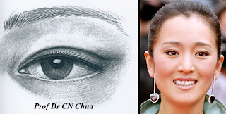
Double eyelids are present in about 50% of Chinese, however, their shapes are distinctly different from that of Caucasian. The types of double eyelids seen in Chinese can be broadly divided into the tapering type (开扇形) and the parallel type (平行型). In the tapering type, the double eyelid is joined to the edge of the eyelid at the nasal (inner) side and becomes progressively higher as it moves temporally (outside). In the parallel type, the double eye (skin crease) runs parallel to the eyelid edge along its full length. A study in China found that tapering type accounts for 80% of Chinese who have double eyelids and the parallel type 20%.


The other type of double eyelid which is rare in Chinese but typical of Caucasians is the semilunar type (新月形) in which the double eyelid is highest in the centre and lower at both the nasal (inner) and temporal (outer) corners. Below are some more examples.

Tapering double eyelid

Parallel double eyelid

Semilunar double eyelid

Sunday, November 28, 2010

A Mini Drama During Double Eyelid Consultation aka Oriental look versus Neither-Here-Nor-There

Last week, a teenager girl came with her mother for double eyelid surgery (Asian blepharoplasty). Both were Malaysian Chinese residing in Australia. At the consultation, the girl produced a magazine (see below) and wanted her eyelids to look like the Caucasian model on the front page. Her mother who had been quiet until then objected strongly and said that would ruin her look: "I don't want the neighbour to think I bring home a Filipino maid!" A fierce argument raged between the daughter and mother over the types of double eyelids each wanted until both were in tears. Seeing the situation was getting out of hand and whatever double eyelid I performed would not please either, I advised them to consider the option carefully.


Personally, I think no responsible surgeons would want to create double eyelids which are deep and high for a teenage girl. To illustrate how she would appear let looks at two Taiwanese Chinese celebrities who had had double eyelid surgeries: Tracey Huang (黃露儀) and Hu Yin Meng (胡茵梦).


By looking at the pictures above, one would have problem placing the ethnic origin of the person on the left (Tracey) but the one on the left (Hu) is distinctly Chinese. This is hardly surprising as it is rare to come across Chinese with deep set eyes and cresenteric double eyelids (more on the shapes of different eyelids in my next blog) except for those Muslim Chinese living in Western China (Xinjiang) or the Native Taiwanese who are of Polynesian origin. Any Chinese who has the look of Tracey is said to have a extremely surgical look to their double eyelids, whereas that of Hu appears natural. Below are their pictures before and after the double eyelid surgery.

Hu Yin Meng (胡茵梦)

Tracey Huang (黃露儀)

Saturday, November 27, 2010

How I Removed a Benign Lesion Without Losing the Eyelashes

After seeing those two patients who were bitter about lossing their eyelashes after removal of eyelid lesions, I was referred a patient with a left upper eyelid mass which was thought to be a sebaceous cell carcioma. However, there was no evidence of involvement of the tarsal plate and the eyelashes were all present making it unlikely to be a sebaceous cell carinoma. Bearing in mind the two patients who were as upset over the lost eyelashes as the initial eyelid problem they had, I decided to plan my operation carefully.

A conspicuous mass in the left uppe eyelid.

No involvment of the tarsal plate.

The eyelashes are present.

One certain way of not creating an area of eyelashes loss is to perform a pentagon excision as in my previous blog. However, the procedure would mean the patient needs to wait at least a month before the eyelid will return to its normal shape. So I decided to perform a limited partial thickness excision of the mass sparing those at the lid margin. The lesion was sent for histology and the defect was closed with double sliding flap or the so-called H-flap.

Partial thickness excision of the eyelid mass.

The lid margin was spared.

Two sliding flaps were created to close the defect.

The flaps were bought together to see if they can be joined
without causing eyelid distortion.

At the completion of the surgery.

Post-operatively, the patient was pleased with the result as the eyelashes were not lost and there was no notching of the eyelid.


Apperance of the eye at 10 days.

The eyelashe were of uneven heights, as some them were cut
accidentally during the operation.

10 days after the operation.
Close-up view of the eye at 10-day post operation.


Friday, November 26, 2010

How I Managed the Lost Eyelashes Part 2

The first patient from the previous blog was a 30 year-old woman who had a smaller area of eyelash loss. I suggested to her that I would excise the area of defect using the partial thickness technique. The defect would then be covered with sliding flaps by spliting the grey line (the border that separates the front and back of the eyelid; see picture below).

However, she was reluctant to undergo the procedure as she was concerned about scarring and the duration of recovery. The alternative would be eyelash tranplant using hair follicles from the scalp but I have never performed this procedure. Either way she was reluctant to undergo any surgical procedure and asked if I were able to give her something that could make the eyelashes grow again. When I looked at the defect closer, I noticed she had three eyelashes in the area of the defect (red arrows showing the eyelashes at the area of defect).

So I prescribed her a bottle of xalatan (latanoprost) and advised her to apply the drop to the area of the defect every night. Xalatan which is used for glaucoma has long been known to make the eyelashes grow longer and darker. Although it may not cause the eyelashes to regrow, I hope it may make the remaining eyelashes to grow thicker and darker and mask the area of the defect. Xalatan has the same effect but cheaper than the recently launched product LATISSE® (bimatoprost ophthalmic solution) 0.03% for eyelash growth.


I shall review the patient again in four weeks and report any changes in this blog.

Thursday, November 25, 2010

How I Managed the Lost Eyelashes

The second patient was a man in his 30s and had a bigger area of eyelash loss. There was also noticeable notching in the defect. I perfomed a pentagon excision to remove the defect as shown below.


Post-operatively, the left eye was droopy for about 3 weeks before returning to its normal height.


The following picture showed that the area of eyelash loss is now removed.


I shall mention how I manage the other case tomorrow.

Wednesday, November 24, 2010

My Eye Surgeon Did That to My Eyelashes

Recently, I encountered two unhappy patients who had had benign lumpy lesion from their upper eyelids resulting in localized loss of eyelashes. When they complained to the eye surgeons (two different doctors) who did the operations, they were reassured that the eyelashes would grow back. However, 6 months later the areas remained barren. When they returned to the eye surgeons, they were told it was not big deal to lose a bit of eyelashes as they would not affect their visual functions. Unfortunately, their grievances were quite justified as the areas of lash loss was easily noticeable. This was more so for the second patient than the first one as the areas of loss in the later was bigger and extended beyong the cornea where the colour of the iris would have masked the defect.

Case 1


Case 2


Examination showed scarrings of the lid margins. The eye surgeons in their enthusiatic attempts to remove the lesions which were not cancerous had caused damages to the eyelash follicles. Although often ignored, eyelashes are important in protecting the eyes from dust and tear which can irritate the eyes. The second patient actually experienced constant discomfort becaue of the size of the defect. I shall discuss how I managed each case in my next blog.

Tuesday, November 23, 2010

Turning Single Eyelid into Double Eyelid

This blog is the continuation of "Live Anatomy of a Single Eyelid" showing how I suture the eyelid to form the double eyelid. As I mentioned earlier, patient with single eyelid the levator muscle (the muscle that open the eye) is attached to the eyelid skin at a lower site compared with those with double eyelids. To create the double eyelid, the muscle need to be reattached at a higher site. To achieve this, the fat in between the skin and the muscles should first be cleared to give good exposure. Only then, the muscle is sutured (or attached) to the skin to form the double eyelid. The pictures were kindly taken by Terence Lim.

The levator muscle is sutured to the orbicularis. This step is
optional but I find it increases the success rate.

The skin is now stitched to the levator at several points to
form the new attachment

At the end of stitchings. The double eyelid appears higher
but this is caused by a combination of swelling and stitching
and will resolve over the next two weeks.

Pictures of this patient will be shown here as she recovers. The following pictures belong to two patients that I did earlier this year using the incision techniques shown here. I will discuss the suture techniques in my future blog. 


Sunday, November 21, 2010

Live Anatomy of A Single Eyelid

This 48-year old patient with single eyelids undergoes a double eyelid surgery. The pictures taken during the operation shows the various structures of the eyelids. The pictures were taken by Terence Lim.

This patiet with single eyelids also has excess eyelid skin.
The second picture shows that when the upper eyelid skins
are lifted the eyes appear bigger.


Fuller single eyelid at the beginning of the operation.

The skin to be removed was marked with a black pen.
The height of the double eyelid is set at 6mm.

The skin is removed to show the orbicularis
(the muscles that shut the eyes).

After removig the orbicularis, the orbital septum comes into
view.  In single eyelid, this structure joins the levator
(mucle that open the eye) at a lower site. As a result, the
fat is situated more anteriorly making the eyelid fuller.

When the orbital septum is open, the orbital fat comes
 forward. Underneath the fat lies the levator muscles.

The fat is clamped and excised.


Once the fat is removed, the eyelid falls backward
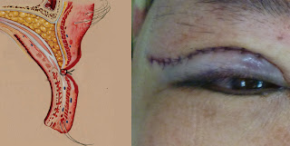
and a double eyelid is formed.

Single eyelid is now transformed into double eyelid.


Saturday, November 20, 2010

Why Some Chinese Have Single Eyelid and Others Double Eyelid?

In the previous blog, I looked at the anatomical differences between East Asian and Caucasian eyelids to explain the absence and presence of doube eyelids (skin reases). Now we look at why some East Asians have single eyelids and other have double eyelids. Again this can be explained by the insertions of the levator muscles (the muscles that open the eyelids) into the eyelid skins. In East Asians with single eyelids, the levator muscles are inserted into the skin either weakly or very low near the eyelashes. Whereas those with doube eyelids, the muscles are inserted strongly and higher above the eyelashes. To demonstrate these anatomical differences, Dr Ngo with single eyelids and Dr Brian Teo with double eyelids kindly agree to be the models.


The low insertion of the levator explains
why Dr Ngo has single eyelid.


The higher insertion of the levator explains
why Dr. Teo has double eyelids.

Friday, November 19, 2010

The Unique Features of East Asian Eyelids

The absence of double eyelids (skin creases) is almost exclusive to the East Asian races (Chinese, Korean and Japanese). It is estimated that 50% of East Asians are born without double eyelids (ie. single eyelids). In the other 50%, the double eyelids are usually indistinct or lower than that of other races. There have been several studies looking into the anatomy of the East Asian eyelids to explain why double eyelids are absent, indistinct or low.


Most of these studies compared the East Asian eyelids with that of the Caucasians.


In East Asians, the muscles that open the eyelids (levator palpebrae) are inserted into the skin of the eyelids at a lower site than in the Caucasians. Depending on the site and amount of insertions, the double eyelids may be absent, indistinct or present. In addition, the fat compartments in East Asians are more protruded which explains a fuller upper eyelids. In Caucasians (and other races), the muscles that open the eyelis are inserted into the skin at a higher site and more firmly so that their double eyelids appear higher and more distinct. The fat compartments in Caucasian are confined to a more posterior position whcih accounts for their eyes appearing deeper and sunken.

East Asian eyelid anatomy.

Caucasian eyelid anatomy.