Thursday, February 27, 2014

A Patient with a Rapidly Growing Eyelid Tumour

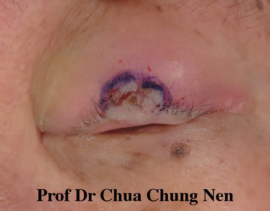
Malignant (cancerous) eyelid tumour is less common in Malaysia than in Western countries because the additional melanin in the skin of the locals protect them against the cancer-causing effect of ultraviolet light. Nevertheless, basal cell carcinoma remains the most common eyelid tumour seen in my oculoplastic clinic. This lesion typically affect people who have chronic sun exposure such as labourers. This 50 year-old man presented with a mass in the right upper eyelid. The lesion first appeared 9 months earlier but grew rapidly along the upper eyelid margin affecting his vision. It also bled easily. Rapid growth and bleeding are two signs suggesting the lesion was a canceroud growth. A biopsy confirmed it was a basal cell carcinoma.

A growth along the right medial 2/3 of the right upper eyelid.

The main objective of treating any cancerous skin lesion is to excise the lesion completely. In this patient, one is faced with the options of either excising the skin tumor alone or take with it 2/3 of the upper eyelid. With the first option, one only need to replace the lost skin but risk leaving behind some tumour; with the second option one can be more certain all tumour is removed but this would leave a big gap needing a bigger reconstruction with longer recovery time and less aesthetic result. In this patient, the tumour invaded only the front of the eyelid with good clear lid margin ( picture b) so I opted for skin excision alone.

Excision of the lesion followed by reconstruction using forehead skin.

The excised lesion was sent for histopathology and after the area was tumour free the forehead skin was used to cover the defect.

Result at one week (pictures a with eyes open and closed) 
and two week (b) postoperative.

The patient was able to open his eye at one week and by two weeks the scars were much improved with steroid ointment, sunscreen and sunglasses. He was unable to return for regular visit as he lived 5-hour drive away. He was advised to return in three-month time to check for any recurrence.

Tuesday, February 25, 2014

An Old Man with Droopy Eyebrow

This 62 year-old man was referred for droopy eyelid from the polyclinic. Examination revealed that the right pupil was covered by the upper eyelid skin which interfered with his vision especially during driving. The past history revealed that he suffered from right facial paralysis two years earlier. Although most of the function of the facial muscles have now returned, the right upper forehead muscle remained severely affected. This has resulted in droopy eyebrow which in turn caused the eyelid to droop over the right eye.

Right eye covered by the upper eyelid skin because of eyebrow droop.
 The picture on the right shows the right forehead muscle is non-functioning
 due to previous facial paralysis.


There were two solutions to the problem: either the excess eyelid could be excised  or the eyebrow could be lifted to open the eye. If the excess eyelid were excised, the eyebrow would droop further and aesthetically the result is poor. The best solution was to lift the eyebrow which would also balance the height of the two eyebrows to give an aesthetically pleasing result.

Steps in lifting the eyebrow directly. a. The height of the droopy eyebrow is lifted 
to the same level as the normal side; b and c. The amount of skin that needs to be 
excised to achieve this is drawn; d. the height is rechecked; e. the skin is excised 
and f. closure of the defect lift the eyebrow, initially the amount of lift is more 
than expected due to suture tension this would loosen over the next few days. 

Good height at one week postoperative.


Sunday, February 23, 2014

A Severe Case of Out-turning of the Lower Eyelid

This 70 year-old man had been suffering from right watering and sore eye for the past year. He saw a few GPs who only prescribed topical antibiotic but the condition did not improve. Eventually he consulted a different GP who recognised his problem to be ectropion ( a medical term for out-turning of the eyelid) and referred him to my oculoplastic clinic. 

Watering and red eye from out-turning of the lid.

There are several causes of out-turning of the eyelid but the most common cause is ageing changes in which the eyelid structures become loose. This gives rise to constant watering as the tear can not be held in correct position. Discharge is also common due to constant irritation. The treatment of choice is with surgery to reposition the lower eyelid. There are several methods to choose from depending on the severity and the extent of the out-turning. In this patient, the out-turning extent the whole length of the lower lid so I chose the Lazy-T method to reposition the lid. The Lazy-T is so-called because it involves a vertical and a horizontal cut resembling a letter T lying on its side.

Lazy-T technique in a nutshell.

Steps of Lazy-T technique: a. marking is done before the operation; b. a diamond shape excision is done on the tarsoconjunctival side just below the punctum; c. suture is used to appose the tarsoconjunctival wound and invert 
the punctum; d. a vertical incision is made involving the full length of the tarsus; e. the two ends of the cut lid are overlapped to determine the amount of eyelid to be removed; f. once determined the excess eyelid is removed as a pentagon; g. after the excision; h. the two ends are apposed; i. the tarsus are sutured using 6/0 vicryl and j. the skin 
are sutured with 6/0 ethilon.

Below is a patient I did earlier. She had out-turning of the eyelid involving the medial side of the lower eyelid. A lazy-T technique was used.

A 30 year-old woman with out-turning of the lower lid (top picture) 
and one month after lazy-T technique (bottom picture).


Saturday, February 22, 2014

Another Case of Severe Eyelid Retraction due to Thyroid Eye Disease

This patient has thyroid eye disease for the past 3 years and in the past year had had problems with shutting her left eye. This was caused by severe retraction of the upper eyelid muscle. Despite using regular eyedrop the eye was sore and red. In addition, the vision was severely affected due to cornea changes from dry eye. 


There are several ways of correcting the retraction. In my previous blog I did an upper eyelid recession for eyelid retraction secondary to thyroid eye disease. This technique was easy to perform but the postoperative swelling takes about 10 days to 2 weeks to resolve. For a faster recovery, only the Muller's muscle that open the eye may be excised. This technique cut through less tissue so the swelling tends to resolve faster, however, the lowering effect may not be as much as the technique of upper eyelid recession.

The technique of mullerectomy for eyelid retraction.

Eyelid appearance pre and two weeks post-operative.


Thursday, February 20, 2014

Excision of an Upper Eyelid Lesion followed by Reconstruction

Many skin lesions can affect the eyelids. It is important to differentiate between the benign and the cancerous types as the amount of tissue to be removed differ. In the case of benign lesion, normal looking tissue does not have to be removed whereas in cancerous lesion some normal looking tissue are usually removed as the tumours may invade adjacent tissue without visible changes to the naked eyes. This patient presented with a rapidly growing upper eyelid lesion of one year duration. However, the lesion showed no ulceration or bleeding. To avoid unnecessarily removing a large amount of normal tissue with resultant longer time of recovery after reconstruction, a bit of the tissue was removed and sent for histology before embarking on surgery. This was reported as a warty lesion with no malignant changes. As  the lesion was unsightly and interfere with his eye opening, the patient wanted it removed.

I removed the lesion from the front of the eyelid only instead of doing a full thickness skin excision. Otherwise, the patient would need a larger reconstruction and not be able to open his eye for at least two weeks. The defect of the skin lesion was covered with flap from the loose skin of this patient's upper eyelid.

An eyelid lesion involving the upper eyelid.

An eyelid lesion involving the upper eyelid.

The edge of the lesion was marked.

The lesion was excised from the front of the eyelid.

After the excision.

The intended flap was marked.

The flap was reconstructed.

The defect was closed with the flap.

At the end of the surgery.

The appearance at one day post-operative.

The appearance at 10 days postoperative.