Friday, December 31, 2010

Botched Double Eyelid Surgery in Beauty Salon

This 50 year-old woman presented with bilateral droopy eyelids and unequal double eyelids. She underwent double eyelid surgery in a beauty salon two years ago. Suture technique was employed. She noticed the two eyelid were not right the next day and returned to the beautician for advice. However, she was told that her eyes were normal. Since the operation, she has been reluctant to go out for fear of being ridiculed.


In order to correct the problems, the double eyelids need to be re-operated using the incision technique to lower and balance the double eyelids. It is illegal for non-trained personnel to perform medical procedure in Malaysia, unfortunately, the law is not well enforced. Consequently, there are many botched surgery being performed in beauty salons for quick profit. I shall show more of such cases in future blogs.

Monday, December 27, 2010

Case 2: Excision of Basal Cell Carcinoma with Reconstruction

This patient has a lesion on the nasal side of her right eyelid which bled easily on contact. A biopsy confirmed basal cell carcinoma. Fortunately, the lesion was a distance away from the punctum (the holes that drain the tear) otherwise there was a risk of the tumour spreading into the deep structure. The resulting defect was covered using rotation flap.


A lesion on the nasal side of the right eye.

The tumour was marked with 3mm margin to ensure sufficient
excision of the tumour without leaving behind any tumour cells.

The defect was covered with a rotation flap.

 
The rotation flap was created to cover the defect.

The defect was partially covered by the flap. The remaining
defect was closed directly.

At the conclusion of reconstruction.

Diagrams showing the principle of rotation flap.


Saturday, December 25, 2010

Don't Worry its Just a Small Black Head - An Unwanted Side-effect of Suture Technique for Double Eyelid

This 46 year-old woman underwent double eyelid procedure in a beauty saloon 2 year ago. The procedure was performed by a so-called "beauty surgeon" employed by the saloon. Although, she got her double eyelids but she was unhappy with the excess skin hanging over her skin creases (double eyelids) which the beauty surgeon was unable to do anything. Recently, she had been bothered by constant irritation in the left upper eyelid and a black spot. She returned to the beauty saloon but was told by the owner that it was just a small black head and there was nothing to worry about. When the patient asked to see the beauty surgeon, the saloon owner says she had no idea where she was as she only employed those surgeons on one-off basis.
Poor double eyelid formation because the excess upper
eyelid skin was not addressed.


A black spot causing recurrent irritation in the left
upper eyelid.

When I examined her the black head appears to be the end of the suture used to form the double eyelids. The "black head" was cut and removed it in entirety. 

The suture was pulled and cut.


The whole suture removed and appeared to be 6/0 nylon.

Although suture technique is useful in young patients,  in older people with excess upper eyelid skin (dermatochalasiss), it does not work well. The treatment of choice for this lady would be to combine double eyelid surgery with removal of the excess skin and this could be achieved with incision technique.

The suture technique used in this patient is the two-way continuous buried-suture approach as shown in the diagram above. Picture A 7 to 9 incisions for the creation of a pretarsal crease are made with the tip of a No. 11 scalpel blade, approximately 1 mm long, and are placed approximately 5–7 mm apart in a gentle curving arc. The line begins 5 mm lateral to the medial canthus and ends 10 mm lateral to the lateral canthus. Picture B The first pass of the suture is from lateral to medial, capturing dermis between alternating incisions and superifical tarsal plate between dermal bites. Picture C The suture is then reversed and passed from medial to lateral, capturing dermis and tarsus in the opposite intervening segments.

Tuesday, December 21, 2010

Ma'am I think you have swallowed the tube

Last Friday, two of my patients who had had DCR (dacryocystorhinostomy) surgery for blocked nasolacrimal ducts returned for removal of the tubes which were inserted to ensure patency of the connection between the tear sac and the nose. The removal involved cutting the tube at the eye level and asked to blow their noses hard to expel the tube.

The first patient was a 50 year-old man who did this without problem.

Tube at the level through the holes that drain tear.

The tube was cut at the eye level.

 The patient was instructed to blow his nose.

The tube that was blown out of the nose.

The second patient was a 75 year-old woman who had problem blowing her nose. So I attempted to expel the tube by flashing the tear duct. Although she could feel the water running down her throat, the tube was not expelled through the nostril. Consequently, I have to examine her nasal cavity for the missing tube. Unfortunately, the tube could not be found. I suspected the tube might have been flushed into her throat and swallowed. The tube is not toxic and should not cause any untoward effect.

The DCR tube at the eye level.

The tube was cut with a pair of scissors.

The cut tube could now be removed through the nostril.

The nose was inspected for the missing tube.

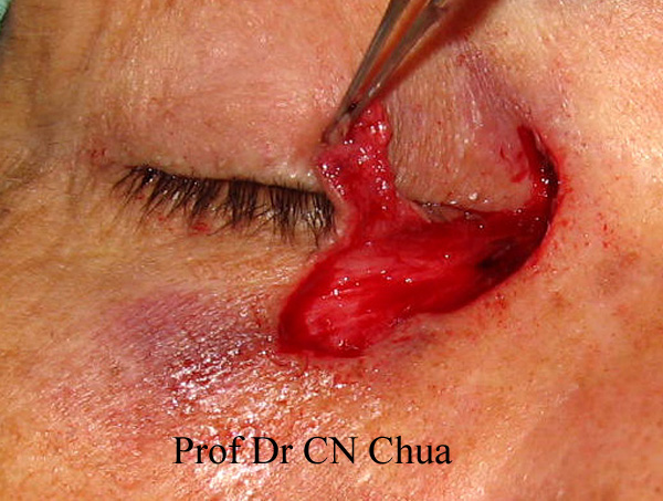
The arrow indicates the hole created to join the tear sac
to the nose so that the tear can be drained.

Diagram showing where the tube might go.

The blue circle indicates the hole (fistula) created between the tear sac and the nose.The green lines represent the DCR tube. The tube usually came out through the nostril (red arrow) when one blow the nose. The tube might have been swallowed because during flushing in the second patient, the tube might have been pushed into the throat (blue arrow).

Sunday, December 19, 2010

Case 1:Eyelid Reconstruction for Eyelid Basal Cell Carcinoma

Basal cell carinoma is the most common type of skin cancer involving the eyelid. It is also known as rodent ulcer because it was thought to be caused by bite from rat. It is associated with excessive ultraviolet exposure and usually occurs in people who work outdoor or enjoy outdoor activities such as golfer. The tumour spread by slowly eating away the surrounding normal tissue and can cause severe destruction if ignored.
This  65 year-old man presented with a lesion in the left medial canthus (area between the nose and the eye). The lesion had been present for the past 9 months and had got progressively bigger and bled easily on scratching. The feature was suggestive of basal cell carcinoma.


The lesion was excised with a 3 mm margin. The histology confirmed basal cell carcinoma with tumour-free margins. The defect was covered with an O-Z plasty as shown.


The area to be excised was marked together with
the pattern of reconstruction.

Principle of OZ plasty.

At the end of surgery.

Appearance at one week post-operative.


Friday, December 17, 2010

People Who Shed Bloody Tear (Haemolacria)

It is not unusual to hear claims of religious statue usually Virgin Mary weeping bloody tear which attracted flocks of pilgrims or curious on-lookers. As often as not, these claims turned out to be hoax as the weeping Mary in Sacramento, California.


However, bloody tear in human being is certainly the most alarming and unusual complaint. Amongst the few reported cases almost all the sufferers were women, therefore, it is unusual for me to come across this boy who is reported to have shed bloody tear for the past 2 years sometimes lasting up to an hour. He is reported to have seen more than 15 eye specialists but no cause could be found.

Alarming bloody tear.

Blood coming down the face and nostrils suggesting that
the nasolacrimal ducts are not blocked.

The differential diagnosis of bloody tear is wide and includes severe anaemia, jaundice, vascular tumours, Osler-Weber-Rendu disease (hereditary haemorrhagic telangiectasia) haemophilia and other coagulopathies, tumours of the lacrimal sac, epitaxis. In this patient, the first line of investigation would be to make sure that the tear is indeed blood and the blood is of the same group as his to exclude a hoax. Detailed eye examination should be performed to look for any possible causes which I am sure his previous eye surgeons had performed. It would be interesting to hear what the final diagnosis is. His case is being made into a documentary and will appear in The Learning Channel.

Wednesday, December 15, 2010

Common Presentations of Eyelid Cancers - the Straight Forward Cases.

As the only oculoplastic surgeon in Sarawak (population 2.5 millions), I have the opportunities to see a variety of eyelid cancers. Fortunately, most of the cases could be removed with straight forward recostructions that don't have to test my surgical skills to the limit. Here are a selected cases I came across in the last year. All of them show the same features of tissue destruction with easy bleeding and loss of eyelashe. I shall show the techniques I used to reconstruct the tissue defects. However, it is important to bear in mind that as my oculoplastic teacher Miss Jones used to say at the end of the day no matter how good the recontruction is the complete removal of the tumour should take precedence. More complicated eyelid tumours will follow in the future blog.

Case 1: A 65 year-old man with a skin lesion with recurrent
bleeding.

Case 2: A 62 year-old woman with a nodular lesion and
easy bleeding on rubbing.


Case 3: A 72 year-old man with a left skin ulcer and recurrent
watering eye. He had had excision performed one year ago
in another centre and the report showed the tumour, basal
cell carcinoma was not completely removed.


Case 4: A 58 year-old man with a large dark nodule which
bled easily.


Case 5: A 61 year-old man with a left lower eyelid cancer.


Case 6: A 53 year-old man with a left lower eyelid skin lesion
that failed to heal despite antibiotic.


Case 7: A 79 year-old man with a left lower eyelid white patch
with loss of eyelashes.A biopsy in a private centre showed it
to be squamous cell carcinoma.

Tuesday, December 14, 2010

Impressive Surgery to Transform Facial Blemish

Came across this interesting piece of news in UK Daily Telegraph then went to the Chinese website to search for more. The skill of the surgeons is making me jealous, wish they can share their experience by publishing the case in Scientific Journal but so far can't find it on-line in either English or Chinese.

PhD student Tao after facial plastic surgery to remove a massive facial mole, left, taken on Sept. 23, 2009 and before the surgery, right, taken on March 24, 2008, both in the hospital in Hangzhou in east China's Zhejiang province.

Below are furher pictures and report from Chinese website, there is no mention where the skin was taken from but I presume it would be either the buttock or the inner thigh for a combination or full and split skin grafts.

“黑脸”博士变脸白面书生 想要事业爱情双丰收


    一块兽皮一样的巨大黑色痣从左脸蔓延过鼻子,又延伸到右侧颧骨,昨天,记者见到24岁的缙云小伙子小陶容貌特别,250平方厘米大的巨型黑痣盖住了他本来端正的眉眼。“我明年博士毕业就出国了,以后还要找女朋友,容貌好了生活上会比较方便。”小陶是浙江大学在读博士,今天上午,他将在杭州时光医疗美容医院接受“面部巨大黑痣切除并植皮术”。


  “我能坦然接受这样备受关注的目光,能理解。”昨天上午,记者在杭州时光医疗美容医院病房里见到小陶。面对几个一直盯着看的记者,一开口,小陶就流露出幽默感。其实记者们关注的,除了他脸上的巨型黑痣,还有他本科双学位、浙大博士的传奇经历。

  巨大黑痣盖在脸上,照常推理,即使是男孩子也会介意,本以为小陶跟多数人一样,因为容貌影响而自卑、自闭甚至拒绝与人交往,急需通过整形重树自信人生。聊了几句,几个记者才领略到,容貌特别的才子小陶,开朗阳光,笑容自信,侃侃而谈的一副学者模样。


       “这块痣对别人来说可能是自卑的根源,但对我来说,可能是上进的动力。太在意它只会徒增烦恼啊!”记者的问题都集中在小陶的脸上,他一一从容回答,偶尔开玩笑,自己先笑了起来。1984年出生在浙江缙云的小陶,在爷爷奶奶的辛苦抚养下长大。
     
      “乡下人对我的脸说法很多,有的人说这是非同一般的人,我虽然不完全相信,但还能接受。不过现在想想,那个闭塞的小乡镇,给我很多宽容。”小陶说。


       学业紧张没时间做手术

 从小学到高中,小陶几乎没有受过歧视的目光,学业出色的他得到同学更多的尊重。“如果算是挫折,就是高考的时候吧。招生老师看到我档案上的照片,让我连夜赶到杭州面试,最后6位老师一致觉得一定要把我招进去。”
勤奋好学的小陶,在大学时曾动了整容的心思,可是要住院一个月,他想了想,白天晚上都上课,整形只好给学业让路,就耽误下来。


    容貌特别的才子小陶,本科毕业时,拿到了物理专业和数学专业双学位,并顺利推荐保送到浙江大学理学院硕博连读,据说当时,成绩优异的他“一封Email就把自己成功推荐给了现在的导师”。“我的研究方向是生物矿化,打个比方就是让牙齿脱落后重新长出来。我明年毕业,现在已经发表了十几篇学术论文了,打算赴美继续深造。”

  整形后有望貌似潘安

  小陶偷偷告诉记者,他想整形,一部分原因是找女朋友。“我跟以前的女朋友感情很好,她也并不介意我的黑脸,可是,等到想见家长时,压力就大了,后来我们分手,我想主要原因是黑痣的问题。”小陶是个蛮重感情的小伙子,他说,找女朋友光有自信和人格魅力还不够,如果去掉黑痣,交女朋友顾虑就会少很多。已经过世的爷爷奶奶曾经有个遗憾,没能见到长孙去掉黑痣的样子。

  “说起来凑巧,我有一次逛植物园,一个不认识的大伯,说有个小女孩跟我一样的痣,在杭州一家医院治好了,还建议我去打听一下。后来我陪老家来的表弟治疗太田痣,就到了时光医院,表弟解决了问题,我也动心了。”

  昨日,小陶接受了全面的术前检查,医生表示,手术技术已经非常成熟,小陶的眉眼本来就很端正,才子小陶整形以后可能“貌似潘安”。


       先天性巨痣是黑色素细胞痣的一种,为较少见的一种先天性皮肤良性肿瘤,其特点是范围广泛,手术切除后的组织缺损往往不能为邻近的局部组织所修复,尤其是位于颜面部的巨痣,因影响外貌美给患者带来严重的心理障碍,其发病率接近万分之一。

        “黑面”的他阳光自信

Sunday, December 12, 2010

Antique Prints Showing Eyelid Cancers

Eyelid cancers have been known since ancient time, however, due to the lack of medical knowledge coupling with primitive medical practises most of these cases just grew and grew causing significant facial disfigurement. Many of these patients eventually died from the diseases. Here are four antique prints showing eyelid cancers from the Wellcome Trust in London.
A Chinese woman with a large left eye tumour
which covered the whole eye. Cause unknown.

A tumour arising from the eyelid which grew
to cover the left eye. The exact cancer is unknown.

The left eye is covered by a fungating tumour
said to be an osteoma.
A basal cell carcinoma (rodent or Jacobi ulcer)
had eaten away the tissue in and around the
right eye

Friday, December 10, 2010

Would She be Able to Open Her Eye Again?

The patient in my last blog returned the next day for review. The reconstruction appeared secured but the family was concerned that she may not be able to open her eye again.

24 hours after excision of tumour and reconstruction.
The upper eyelid is closed.

So I have to reassure them with pictures of patient (see pictures below) who had had similar reconstruction. This 50 year-old man had had recurrent left upper eyelid stye which was repeatedly incised and drained. In the last treatment, the surgeon sent the specimen for analysis which showed sebaceous cell carcinoma. This tumour need early removal as it may spread to other parts of the body and cause death.

Left upper eyelid tumour (see picture above).  The marking on the eyelid showed the amount of tissue to be removed and the marking on the side of the eye indicates the amount of tissue needed to cover the defect during reconstruction.

The tumour was excised with 5mm clear margin to avoid recurrence (see picture above). The eyelid was closed using the technique of reversed Tenzel's flap by mobilizing the tissue on the temporal side of the eyelid.


The left eyelid could not open immediately after the operaton. However, the left upper eyelid returned to near normal appearance at 4 weeks follow-up.