Thursday, February 25, 2016

Doc, Do I Need Operation on My Epicanthal Fold (aka epicanthoplasty, 開眼角) ? - Part 5

There are over 30 methods described for the correction of epicanthus. Some are more complicated than the others, however, the results of the complicated ones are by mean superior than the simpler ones. In addition,  surgery on the inner canthal region is prone to skin contracture and scarring and therefore a method that combines the least number of cutting and cut near the lid margin is preferred. 

Some of the epicanthoplasty described are shown below (from Asian Facial Cosmetic Surgery). 

Blair method.

Converse method.

Arlt method.

Hiraga method.

Rogeman mthod.

V-W advancement method.


W-plasty method.

Y-V method.


Mustarde method.


Park Z-plasty method.


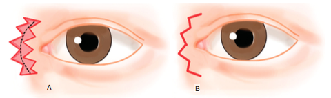
Correction of type IV epicanthal folds in blepharophimosis. The picture on the left 
was done with Y-V method showing minimal scarring whereas the one shown on 
the right was corrected with Mustarde method showing significant skin 
contracture and scarring. Pictures taken from an article discussing
the various methods of epicanthoplasty.


My preferred methods for type II and type III epicanthal folds are Park-Z plasty and skin redraping methods. The later will be described in the next blog.

Type III epicanthal fold corrected with Park-Z epicanthoplasty.

Sunday, February 21, 2016

Doc, Do I Need Operation on My Epicanthal Fold (aka epicanthoplasty, 開眼角) ? - Part 4

Most East Asian babies are born with type III epicanthal folds which gradually change to type II as the nasal bridges develop. Some children with prominent epicanthal folds may be misdiagnosed as having squint this appearance usually improve as the epicanthal folds (from Type III to II) evolve to reveal more of the eye white.


A boy with suspected squint turned out to have epicanthal folds covering the eye white.
 This is also called pseudo-strabismus. By pinching the nose, the eyes appear straight.

In some adults, probably due to low nasal bridges, the type III epicanthal folds can give rise to persistent squinting appearance. Operation on the epicanthal folds not only lengthen the eyes but remove this squinting appearance.

A patient with type III epicanthal folds giving an appearance of squinting. 
Epicanthoplasty was done together with filler rhinoplasty.

Saturday, February 20, 2016

Doc, Do I Need Operation on My Epicanthal Fold (aka epicanthoplasty, 開眼角) ? - Part 3

As mentioned in the previous blog, the medial canthus can be classified into four types depending on the presence and absence of epicanthal folds and the types of epicanthal folds. Obviously, people with type I do not need epicanthoplasty, whereas type II to IV have variable amount of epicanthal folds which can benefit from epicanthoplasty. However, the effect of epicanthoplasty on the width depends very much on the extent of the epicanthal folds with the results being most obvious in type III and type IV (see picture below). 

The pictures show the pre- and postoperative epicanthoplasty (+ double eyelid surgery 
in type II and III). The most obvious changes to the width of the eye are most noted 
in type III and IV.

In the rare cases of blepharophimosis with type IV epicanthal folds, the patients often appear to have a less intelligent facies then they actually are. Many of these patients got bullied at school for their appearances, early operations to lengthen the eyes and correct the ptosis can have a dramatic effect on the patients' well-beings.

The above pictures show a 7 year-old girl who underwent blepharophimosis 
correction with epicanthoplasty and frontalis slings.

Friday, February 19, 2016

Doc, Do I Need Operation on My Epicanthal Fold (aka epicanthoplasty, 開眼角) ? - Part 2


The medial canthus can be classified into four main types type I to type IV (see pictures below) depending on the amount and type of epicanthal fold present.  Amongst the East Asians, type II and type III are the most common (with type II being more common than type III in adult East Asians) accounting for more than 90% of cases, type I is present in less than 10% of East Asians. Type IV is rare and is usually a pathological condition seen in congential anomaly such as blepharophimosis.


Type I: Full exposure of the lacrimal caruncle (in red). There is no epicanthal fold in this type. This is seen in most Caucasian people.

Type II: The lacrimal caruncle is partially covered, the epicanthal fold joins the skin at the margin of the lacrimal lake. This is the most common type amongst adult East Asians.

Type III: The lacrimal caruncle is covered almost completely, the fold in the lower eyelid curves laterally to blend in with the lower eyelid skin. The medial palpebral fissure appears round. This type of epicanthal fold is most commonly seen in young East Asians but as the nasal bridge grows, the epicanthal fold gets pulled progressively to the centre to become type II epicanthal fold. 

Type IV: This is rare and is a reversed epicanthal fold. The fold originates from the lower eyelid and blends with the upper eyelid skin, this type of epicanthal fold is the most severe and make the eye looks exceedingly small.

Using the classification above, you may like to try classifying the epicanthal folds in the following faces (answers at the bottom)


a. Type II without double eyelids
b. Type IV a patient with blepharophimosis, note the small eyes and droopy eyelids.
c. Type I with double eyelids.
d. Type II with double eyelids.
e. Type III in a young girl.
f. Type II without double eyelid.


Wednesday, February 17, 2016

Doc, Do I Need Operation on My Epicanthal Fold (aka epicanthoplasty, 開眼角) ? - Part 1

Many patients planning to have Asian blepharoplasty (double eyelid surgery) often ask if it is necessary for them to also have simultaneous epicanthal fold operation (aka epicanthoplasty, 開眼角). The answer depend very much on what the patients want, if the patient just wants upper eyelid creases then double eyelid surgery is all that is needed. However, if the patients want their eyes appear longer or less squinting (as in some patients with significant epicanthal fold which will mention later) then epicanthoplasty can be performed. 

While epicanthoplasty may look good on patients, it is important to bear in mind some possible disadvantages:
a. Epicanthal fold is characteristic of East Asian eyes, excessive removal may alter the ethnic appearance of the patients;
b. While epicanthoplasty may make the eyes longer, the two eyes will also appear closer together;
c. The area around the epicanthal fold is thicker and more easily scar, so it is important to find an experienced surgeon. 

Before discussing different methods of epicanthoplasty, it is worth taking a look at epicanthal fold. The epicanthal fold is made up of a fold of skin that extends from the upper eyelid across the medial part of the eye to the margin of the lower eyelid. This structure is seen in foetuses of all races before birth, however, except in the East Asians it usually disappears before birth. This structure is seen in over 90% of East Asians origin but only in 2% of Caucasians. 

The epicanthal fold covers the lacrimal caruncle (the red fleshy part of the medial part of the eye) and therefore cover part of the eye. This is one of the reasons why East Asians appear shorter than other races.

The black arrow in the upper picture denotes the epicanthal fold which covers 
some of the lacrimal caruncle (LC) as compared with a Caucasian eye 
which has no epicanthal fold and the lacrimal caruncle (LC) is fully exposed.


The effect of removing or modifying the epicanthal fold on the length of the eye is best demonstrated in the pictures below:

The picture above shows a patient with epicanthal fold and the length of the eye 
is denoted as a. However, when the epicanthal fold is removed (in this case 
by moving the fold digitally) the length of the eye is increased (a +b) through
 exposure of the inner corner of the eye and the lacrimal tubercle (b). 
The effect on the length of the eye depends on the extent of the epicanthal fold.


Monday, February 15, 2016

Doc, Could You Balance My Eyes for the Festive Season? - Part 3

The patient returned on the sixth day of the Chinese New Year for correction of the ptosis. A posterior approach conjunctival mullerectomy was performed.

Pre-operative appearance showing left ptosis.

The pictures below show the steps taken during the ptosis correction:

a-d. Excision of 10mm of conjunctival muller complex followed 
by suturing of the stump of the complex to the tarsal plate.

The principle of the operation is shown diagrammatically below:

Picture on the left shows ptosis due to dehiscence of the levator;
 picture on the right shows creation of a strip of conjunctival muller muscle complex.


Picture on the left shows excision of the strip of the complex leaving behind a stump;
picture on the right shows suturing of the stump to the tarsal plate causes plication of 
the levator muscle thereby elevates the lid.

One hour postoperation showing correction of the ptosis but the eye was still 
droopy due to the effect of the anaesthesia and postoperative swelling.

One day postoperation showing a slight left ptosis correction but this usually 
resolves within two weeks. There is delayed ecchymosis which is common 
in old patients with loose skin and usually disappears within one week.

Appearance of the eyes at 6th day postoperative showing resolution 
of most bruises.


Thursday, February 4, 2016

Doc, Would I Look Ok for Chinese New Year if I Have My Double Eyelid Surgery Done Today?

With the Chinese New Year less than 4 days away, some patients ask to have double eyelid surgery today and hope to look good for the festive season. However, even with the shorter down time of suture technique or small incision, there are too many variables to assure the patients that their eyelids would recover in time. These variables include the differences in vasculatures (blood vessles) of the eyelids (which may mean some are more prone to bleeding and hence bruises) and the susceptibility of the eye muscles to anaesthesia and surgery (so that the eyelids may be droopy for awhile).

While waiting for the surgery, a bit of make-up should do the tricks of brightening up the eyes for the festive season. This technique has been used since time immemorial.

Applying make-up to make the eyes bigger has been in use for thousand 
of years as shown in this picture of an Egyptian Pharaoh. 


The illusion of bigger eyes through make-up is called the Delboeuf's illusion named after Leopold Delboeuf (1831 – 1896), a Belgian scientist. 

Two similar-sized black circles but the one on the left 
appears bigger when it is surrounded an adjacent annulus. 


Nature has also made use of Delboeuf's illusion to make some animals appear cutter. 

Panda with and without the dark patches likened to mascara. 

It is also noteworthy that it is not necessary to apply make-up all round the eyes to make the eyes appear bigger as shown below. 
Single eyelid without make-up.

Eyes appear bigger with make-up around the eyes.

Even a bit of make-up is enough to make the eyes appear bigger. 
Here the make-up is only applied to the central upper eyelids.

Effects of mascara on perceived eye sizes. 


HAPPY CHINESE NEW YEAR


Tuesday, February 2, 2016

Doc, Could You Balance My Eyes for the Festive Season? - Part 2

Although ptosis correction using posterior approach conjunctivomullerectomy has short downtime, the patient refuses to take any chances for fear of lid swelling during the Chinese New Year. As a temporary measure, she is given topical phenylephrine to be used 3hourly. The pictures below shows the result before and after topical application of 2.5% phenylephrine. 

a. Left ptosis; b. After application of 2.5% phenylephrine to the left eye.

2.5% phenylephrine (mydrifin)

Phenylephrine causes lid retraction by stimulating contraction of the Muller's muscle. It has a duration of action of about 3 hours and when taken four times a day should keep the eyelid open during the waking hours. The main unwanted side effects are moderate pupil dilatation which may cause glaring and the risk of precipitating acute angle glaucoma in susceptible patients. The risk of acute angle glaucoma is minimal in patients who have had cataract surgery. 

Diagrams showing the effect of topical phenylephrine. By contracting 
the Muller's muscle, phenylephrine can open the eye by between 2 to 3 mm. 


Monday, February 1, 2016

Doc, Could You Balance My Eyes for the Festive Season? - Part 1

With the Chinese New Year round the corner, most patients want to defer surgery until the festive season is over. This patient is distressed by her droopy eyelid after undergoing a left cataract surgery one month ago in another clinic. Examination shows she has a left significant droopy eyelid (ptosis) and extracapsular cataract extraction and implant (ECCE +IOL). 

Left significant ptosis.

Left eyelid lower when the patient looks down. 

Left eyelid has reduced excursion on upgaze.

Left ECCE + IOL which is likely to contribute to the droopy eyelid.


Nowadays, most cataracts are removed with phacoemulsification (clicked here for video of the technique) under topical anaesthesia, this techniques has the advantages of faster recovery due to smaller wounds and less post-opertive inflmmation compared with ECCE+IOL. Unfortunately, the surgeon this patient went to was not trained in phacoemulsification and still offers the older technique of ECCE +IOL. The droopy eyelid is likely to be the consequence of a longer post-operative inflammation causing dehiscence of the muscle (levator) that opens the eyelid.

Diagrams showing normal levator attachment and following it 


The forehead muscle helps with eye opening. When it is stopped from doing 
so by placing the finger firmly above the eyebrow, the droopy eyelid is worsened.

The eyelid excursion on upgaze is also limited when the forehead muscle 
is not used.

Although droopy eyelid caused by post-operative inflammation may improve when the inflammation settles,  in eyes that develops levator dehiscence the droopy eyelid is unlikely to fully recover. The only permanent treatment is surgical correction to reattach the levator. Posterior approach conjunctivomullerectomy is suggested but the patient is not keen for surgery for fear of post-operative swelling which may not recover in time for the Chinese New Year. However, she is desperate to look her best for the festive season.