This 72 year-old woman from a remote village presented with a one-year history of a rapidly growing lesion of the upper eyelid. The lesion bled easily and contained cheesy substances. Biopsy showed sebaceous cell carcinoma. Untreated this condition can grow rapidly and cause death by either spreading to other parts of the body or destroy the surrounding tissues (see pictures below). In this patient, the upper eyelid was removed and reconstructed using lower eyelid (Cutler-Beard's technique). The flap was left for 6 to 8 weeks to allow formation of new blood supply before being open.
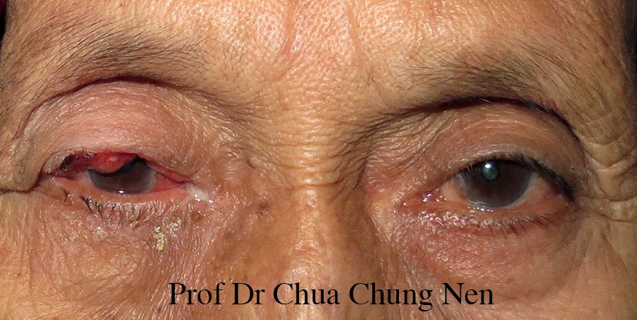
A rapidly growing mass in the right upper lid
destroying most of the eyelashes.

The mass involved nearly all the upper eyelid and biopsy
showed this to be sebaceous cell carcinoma.

A young man who ignored a rapidly growing left eyelid lesion which turned up
to be sebaceous carcinoma until it invaded deeply destroying
most of the ocular tissues.

Another case of sebaceous cell carcinoma which invaded
the brain despite previous surgery.

Steps of upper eyelid reconstruction in this patient. a-c. The upper eyelid
was excised with normal looking tissue to ensure the margin is free
of tumour. d-g. Full thickness lower eyelid was used to cover
the defect. This was done in 3 layers: conjunctiva of lower lid
to conjunctiva of upper lid; orbicularis muscle of lower lid
to levator of the of the upper and skin to skin.
h. End of the procedure.

Appearance of the eye at one week post-operative. The patient was
discharged and given date for opening the flap in 2-month time.
(To be continued)
No comments:
Post a Comment