Many skin lesions can affect the eyelids. It is important to differentiate between the benign and the cancerous types as the amount of tissue to be removed differ. In the case of benign lesion, normal looking tissue does not have to be removed whereas in cancerous lesion some normal looking tissue are usually removed as the tumours may invade adjacent tissue without visible changes to the naked eyes. This patient presented with a rapidly growing upper eyelid lesion of one year duration. However, the lesion showed no ulceration or bleeding. To avoid unnecessarily removing a large amount of normal tissue with resultant longer time of recovery after reconstruction, a bit of the tissue was removed and sent for histology before embarking on surgery. This was reported as a warty lesion with no malignant changes. As the lesion was unsightly and interfere with his eye opening, the patient wanted it removed.
I removed the lesion from the front of the eyelid only instead of doing a full thickness skin excision. Otherwise, the patient would need a larger reconstruction and not be able to open his eye for at least two weeks. The defect of the skin lesion was covered with flap from the loose skin of this patient's upper eyelid.

An eyelid lesion involving the upper eyelid.

An eyelid lesion involving the upper eyelid.
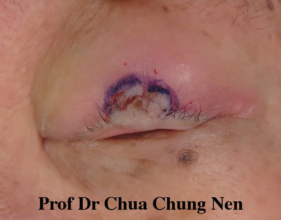
The edge of the lesion was marked.

The lesion was excised from the front of the eyelid.

After the excision.

The intended flap was marked.

The flap was reconstructed.

The defect was closed with the flap.

At the end of the surgery.

The appearance at one day post-operative.

The appearance at 10 days postoperative.
No comments:
Post a Comment