Tuesday, December 26, 2017

Multiple Stones in a Woman's Lower Lids

This 60-year-old woman complained of swelling of the medial lower eyelid with discharge. Examination revealed inflammation of the lower canliculus with pus. The canal oculus was incised and curretaged multiple yellow concretions were expressed. The canaliculus was irrigated with antibiotic and treated with iodine application. This was a case of chronic canaliculitis caused by the bacteria Actinomyces Israeli. 


Friday, December 22, 2017

Social implication of eyelids

Eye contact accounts for 90% of face-to-face communication, it’s little wonder that any imperfection of the eyelids can create a negative impression which can affect one’s social life including career prospect. This 25 year-old insurance agent complained that despite his qualification and selling skills, his clients regarded him as distant and uninterested. He attributed this to his heavy eyelids and narrow eyes which gave him an expressionless face. I created 6mm double eyelids for him with fat removal to reduce the heaviness of the upper lids. The picture at the bottom was taken one-month later, he was pleased with the results and did not return for further follow-up. I assume he was too busy with his clients to return 😀.


Thursday, December 21, 2017

Treatment of sunken upper eyelid without filler or fat injection.

A 23 year-old woman complained of sunken upper eyelids and absence of skin creases (double eyelids) which made her looked tired and older. She had consulted another doctor who recommended filler or fat injection. However, she was concerned that the injection may not last and the associated period of post-injection swellings. I performed bilateral double eyelid surgery with simultaneous posterior conjunctivomullerectomy to widen the eyes and pulled forward the fat to fill the sunken areas. The picture below was taken 2-month postoperative.


Thursday, August 24, 2017

Doc, Can You Show Me Pictures of How You Do Incisional Double Eyelid Surgery?

In my experience, most patients who wanted Asian blepharoplasty (double eyelid surgery) were happy with diagrams detailing the procedure. However, a small group were curious and wanted to see the graphic details. 

This patient had narrow palpebral apertures (width of the eyes) caused by puffy eyelids from fat and absent skin crease (double eyelids).

The desired height of the skin crease was discussed prior to the operation. 
Indentation was made with a paper clip to give an impression how the eyes 
would look postoperatively.

A side view of the right eye with and without indentation shows that the eye 
the skin away from the edge of the eyelid.


The desired height was marked. In this patient the height was 8mm.
Steps of incision procedure: A. The desired skin crease was marked; B An incision was made along the marked line; C. The fat was removed to reduce the puffiness and allowed better formation of the skin creases, to get to the fat (F) two overlying layers had to be removed first: OO = orbicularis oculis the muscle around the eye and OS= the orbital septum. D. The fat was teased out to expose the underlying levator aponeurosis (LP the muscle that open the eye) ; E. The fat was clamped and removed; F. The levator aponeurosis that appeared as white was now fully exposed; G. The levator aponeurosis (LP) was sutured to the remaining orbicularis oculi (OO) anteriorly; H. The skin was closed with 6/0 nylon.

Immediately after the surgery. The asymmetry was caused by swelling 
and reaction to the anaesthesia.

The appearance of the eye 24 hours later.

Two weeks after the surgery.

Three weeks after the surgery. Most swellings have subsided.

Before and after pictures for comparison.


The following are some other patients who had had incision Asian blepharoplasty. 

Before and one month after operation.

Before and two months after operation.

Before and two months after operation.


Friday, August 11, 2017

A hyperthyorid patient with eye protrusion and severe dry eyes.

Hyperthyroidism is associated with ocular changes which can cause eye protrusion and severe dry eyes. This patient developed hyperthyroidism three years ago. Despite medical control of the condition, she noticed her eyes were becoming more protruded with eyelid retraction and frequent tearing that cause blurring of vision.  The tearing was a reflex response to dryness as a result of ocular exposure. In order to reduce the exposure, the lateral aspects of the eyelids were closed surgically (lateral tarsorrhaphy). The operation was performed as followed:

Hyperthyroid patient with ocular exposure due to eye protrusion 
and lid retraction.

Before surgery, the amount of lid closure was determined by closing 
the upper and lower lid laterally.

Before surgery, the amount of lid closure was determined by closing 
the upper and lower lid laterally.

The amount of tissue needed to be removed was marked.

The amount of marked tissue was incised.

The anterior lamella of the eyelid was split to take out the anterior lamella.

The picture showed the tissue removed with the lateral anterior lamella.

The angle of the eyelid was formed with 6/0 vicryl.

The posterior lamella was closed with 6/0 vicryl.

The orbicularis muscle was closed with 6/0 vicryl before the skin 
also with 6/0 vicryl.

Immediately after the procedure.

At two weeks postoperative. The exposure was reduced.


Saturday, July 29, 2017

A Patient with Upper Lid Swelling from Unknown Filler Injection

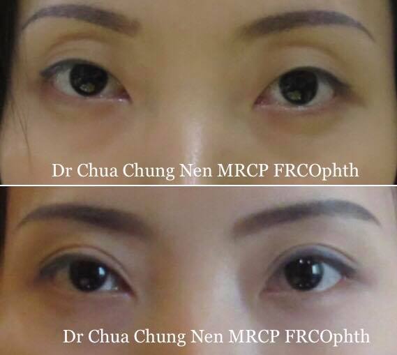
This man had had injection to his upper lid in a beauty saloon with an unknown filler resulting in upper eyelid swellings and occlusion of his visual fields. Examination revealed severe puffiness and hardness of the skin. Hyalase (enzyme to dissolve hyaluronic acid) was injected but the swelling failed to resolve suggesting some sort of permanent filler such as silicone might have been given. 
He underwent upper blepharoplasty with removal of the skin, fat and repositioning of the lacrimal glands.
Severe upper eyelid swelling after filler injection, 
right lid more puffy than the left.

Proposed height of the skin crease was shown to the patient. 
Black arrows shows prominent puffiness of the lateral upper 
eyelids indicating there may also be lacrimal gland prolapse.

The skin to be excised was marked and the skin crease was set 

The marked area was incised. 

The under surface of the excised eyelid showed the filler 
as shinny substances (arrowed).

The orbital septum was removed to show the fat.

The lacrimal gland (1) and levator aponeurosis (muscle that 
open the eye, 2) were found below the fatty tissue.

The fat that formed part of the upper lid swelling was excised.

The lacrimal gland contributed to the lateral puffiness. Removal 
is not recommended as this may cause dry eye.

Instead the lacrimal gland is repositioned using 5/0 ethilon 
to the orbital roof.

The lateral puffiness was abolished after lacrimal repositioning.

At the end of the surgery.


Thursday, July 13, 2017

A Patient with Headache and Blurred Vision.

This patient who had had cataract surgery 5 years ago complained of increasing blurriness of vision and headache. Examination did not show any opacities of the posterior capsule or any retina diseases, however, refraction showed high astigmatism of 2.00 D. In addition, she had excessively high eyebrows. The symptoms were attributable to droopy eyelids: astigmatism can occur when the droopy eyelids press on the globe and the headache due to constant use of the forehead muscle to elevate the eyebrows in an attempt to open the eyes.
After consultation, bilateral ptosis (droopy eyelid) surgery was done using the posterior approach. After the operation, her vision improved due to reduction of the astigmatism to -0.75 and the headache disappeared when the eyebrows returned to normal height.
Headache due to constant excessive use of forehead 
muscle to open the eyes.


One day after ptosis (droopy eyelid) operations.


Normal eye opening two weeks after droopy eyelid surgery.