Monday, November 26, 2012

Correction of Complication of Lower Blepharoplasty 2

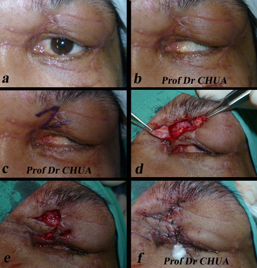
This patient had lower lid blepharoplasty in a beauty saloon two year ago. She complains of dryness of the eyes and rounding of the lower eyelid contour. Examination shows the lower eyelid is tight from excessive skin removal. To restore the normal of the appearance of the eyelid, the skin needs to be replaced. However, the use of skin graft would not aesthetically satisfactory. I recruit the skin by performing disinsertion of the orbiculomalar ligament (see diagram below) and lift the lower eyelid skin by using orbicularis flap (see diagram below) to correct the roundings. 
 
Tightenss of the lower lid and rounding
of the lower lid contours.

Disinsertion of the orbiculomalar ligament.

Disinsertion of the orbiculomalar ligament to recruit
the skin and also to lift the lower lid.

Orbicularis flap is sutured to the orbital periosteum
to lift the lower eyelid.

The picture below was taken at one week after the operation
showing improvement of the retraction
and lower lid contour roundings.

Friday, November 23, 2012

Correction of Complication of Lower Blepharoplasty

Lower eyelid retraction is a common complication of lower blepharoplasty. The main causes are either excessive lower lid skin removal or lower lid laxity.

This 65 year-old man presented with progressive lower lid retraction. He underwent lower blepharoplasty 3 years ago in a beauty saloon. The surgeon was an unlicensed personnel from oversea. He suffered from ocular irritation and recurrent watering eyes. Examination showed severe lower eyelid laxity without significant skin loss.
 
Severe lower eyelid retraction.

Lifting the corner of the eyelid restore the contour of
the lower eyelid. There was no skin shortage.
 
The procedure of choice for this complication is to tighten the lower eyelid as shown in the pictures below.
Steps showing tightening of the lower eyelid using
a procedure known as lateral tarsal strip.

The appearance of the lower eyelids 2 months
post-operative.


Thursday, November 22, 2012

Lower Lid Retraction - A Common Complication of Lower Blepharoplasty


Postoperative changes to eyelid position is a common complication of lower lid blepharoplasty. Most of these cases resolved spontaneously, however, some cases can be permanent resulting in lower lid retraction and rounding of the lower eyelid contour. In addition to the poor cosmetic appearace, these patients can suffer from watering eyes and poor eyelid closure.
 
These complications occur in transcutaneous (in which the skin below the eyelashes is incised) lower blepharoplasty often performed by inexperienced surgeons with removal of too much lower lid skin. Patients at risk are those with lower eyelid laxity when even lesser amount of skin removal can cause lid retraction.
 
The pictures below show different severities of these complications in patients seeking revisional lower blepharoplasty:
 
Mild lower lid retraction with moderate rounding
of the lower lid. The operation was done in a
beautician saloon by unlicensed foreing doctor.
 
Moderate lower lid retraction with
significant rounding of the lower lid. The operation
was done in a beauty saloon by unlicensed doctor.
 
Severe retraction of the lower eyelids with the lower
lids turning out. More severe on the right.
 
Severe retraction of the right lid after transcutaneous
lower blepharoplasty.


Wednesday, November 21, 2012

The Surface Anatomy of an Eyebag

The various structures of eyebags are illustrated through diagram and live anatomy.

The various structures of an eyebag shown through cross section of the lower eyelid.

The various structures of an eyebag as shown through live anatomy of the lower eyelid.

Tuesday, November 20, 2012

What Are the Causes of Eyebags?

I often receive phone calls and emails enquiring eyebag treatments, however, without seeing the patients it is difficult to ascertain what the patients mean by "eyebags". The patients' perception of eyebags may be one or a combination of the following things: loose skin, hypertrophy of eyelid muscle, fat pockets, tear trough, fluid retention to descends of the midface. Therefore, no single anatomic structure can explain eyebags and it is important for the patients to be assessed thoroughly by the surgeon face to face.
The following pictures show pictures of patients presenting with "eyebags":
Eyebags contributed by a = muscles of the eyelid;
b = prolapsed fat and c = tear trough.

Eyebags from tear troughs.

Protruding fats and tear troughs.

Loose skin and descent of the midface.

Protruding fats

Protruding fats and descent of the midface.

Right eye has prominent eyelid muscle
 and the left protruding fat.

Midface descent with protruding fats
and swollen skin on the cheeks (festoons; arrowed)

Monday, November 19, 2012

Lower Eyelid Surgery

Compared with upper eyelid surgery, the lower eyelid tends to be more complicated and if not performed well is associated with more complications. Part of the reasons is the contribution of various anatomical structures that the surgeon needs to address. Not surprisingly, there are many more surgical techniques for the lower than the upper eyelids because of the relative importance of each anatomical structure. It is unlikely that a single technique is sufficient for all aesthetic problems of the lower lid and some inexperienced surgeons avoid doing the lower lid altogether.
 
The different techniques for lower eyelid include:
  • simple fat excision (usually performed from behind the eyelid ie transconjunctival lower blepharoplasty)
  • simple fat excision with lower lid skin excision (transcutaneous lower blepharoplasty)
  • lateral canthal tightening suspension with limited fat and skin excision
  • fat transposition
  • release of retaining structures to abolish the tear trough
  • lower lid bony augmentation with implantation 
Very often the above techniques are used in combination to treat the different aspects of the lower lid blemishes. For example a patient with fat herniation, tear trough and lower lid laxity will need fat excision or transposition, release of retianing structures and lateral centhal tightening.

The photo of this young patient is a good example of the complexity of the lower lid anatomy.  The surgeon need to analyse the importance of each anatomical structure contributing to the lower lid blemish in order to get good aesthetic results.
 
 
a = orbicularis oculis (the muscle under the skin), in some
patient this may be prominent and may be mistaken for eyebag;
b = herniated fat that eyebag usually refers to;
c = tear trough this is a groove marking the attachment
of the muscle to the underlying bony structure.


Tuesday, November 13, 2012

Incision Method for Double Eyelid

Eyelids which are puffy as a result of underlying fat tissue do not do well with the suture technique. The treatment of choice is the incision method as shown in the steps below.
 
This patient has a puffy upper lid (left). To create the
double eyelid (right), incision method was performed.

Before the operation, the desired height was agreed upon by
indenting the upper eyelid with a wire to simulate different
heights of the double eyelid. The desired height
was then makred on the eyelid.

A small amount of skin was first removed, marked a.

Then the orbicularis was removed (marked b), followed by
the orbital septum (marked c) to expose the underlying
 fatty tissue (marked d).

Underneath the fat was the levator aponeurosis (arrowed),
an important anatomy crucial for the success of incision method
and must be exposed along its length. To expose the levator muscle,
 the overlying fat was excised.

The exposed levator aponeurosis (arrowed) was sutured to
the orbicularis muscle along the inferior cut edge.
 
The skin was then closed using interrupted sutures.
 
The incision method causes more swelling than the suture method.
However, most swelling should settle by two weeks.


Monday, November 12, 2012

One of the Many Suture Techniques for Double Eyelids

There are many different ways of doing suture techniques, the one shown here is one such method using three 6/0 nylon sutures.
 
Diagrams showing the placements of the sutures.

Steps in suture techniques:
a. 4 incisions are made on the eyelids.
b, c & d. A curved needle with 6/0nylon is passed full
thickness through one incised area and out through another;
e. The needle is then passed partial thickness through the skin
from the exit site to where the needle initially entered;
 f & g. The suture is tightned;
h. 2 more sutures are similar passed through the lids.


Before & after using the above technique.
 
Before & after photos using the above technique.
 

Thursday, November 8, 2012

A Patient who Can’t Close His Eye after Road Traffic Accident.

This young man was involved in road traffic accident in which he was a motorcyclist. There was substantial loss of eyelid skin and he was referred to the plastic surgeon who performed skin graft using post-auricular graft. Unfortunately, there was shrinkage of the grafts resulting in upper and lower eyelid contracture. The patient was referred to my clinic for further treatment. On examination, the patient had severe lower eyelid contracture resulting in eversion of the lower eyelid (cicatricial ectropion) which interfered with eyelid closure and caused constant watering.
 
Severe eyelid contracture interfering with lid closure (right).
Arrows show shrunken grafts.
 
As there was also skin shortage in the upper lid, I performed a nasojugal flap. He was able to close his eye better at 3-week follow-up.
 
Marking of the flap before surgery (left) and at the end of surgery (right).
The eyelids were tied together for one week to minimize flap contracture.

Diagrams showing how the procedure was performed.

Right picture shows the appearance at one-week
post-operative; left three-week.
 

Tuesday, November 6, 2012

A Patient with Watering Eye after Road Traffic Accident

Malposition of the eyelid(s) following road traffic accident is a common referral to my eyelid clinic. Most of these patients are motor cyclists.
This patient complains of discomfort and watering in his left eye one month after motorcycle accident. Examination reveals skin contracture and out turning (ectropion) of the lower eyelid. CT scan showed compounded fractures of the facial bones. The eyelid appearance is caused by a combination of skin loss and tissue entrapment in the fractured bones.
Contracture of the lower eyelid skin causing eversion
of the lower eyelid.
There are several methods of correcting the malposition. The method I employ is a rotation flap using the skin from the upper eyelid. The discomfort and watering are significantly reduced at one month follow-up. Further surgery may be needed later to reconstruct the facial bony fractures.
a. Marking of the upper lid; b. Release of the lower lid skin scar
c. Creation of the rotation flap; d. Rotation flap in place
e. End of th surgery and the lids are closed together
to prevent skin contracture; f. Appearance the second day.


Improved lid position at 4-week follow-up.

Sunday, November 4, 2012

A Patient with Problem Closing His Eye after Road Traffic Accident

This motor biker was involved in a road traffic accident resulting in loss of his upper eyelid skin. The repair performed in the emergency room did not replace the lost skin and the patient was left with difficulty in closing his left eye which gave him constant eye pain and blurred vision.
Left upper lid contracture from skin loss resulting
in closure problem.
He was referred to the eye clinic for further management. On examination, there was skin shortage resulting in left upper lid skin contracture. I performed a Z-plasty to lengthen the eyelid vertically and the lid closure improved.

Steps taken to correct the contracture.
a. and b. before the operation.
c. marking of the z-plasty;
d. creation of the z-plasty and release of scar tissue;
e. the contracture released with good eye closure;
f. at the end of the procedure, the upper and lower
eyelids were tied together for five days
 to prevent skin contracture.
 
 
3 weeks after the operation. The upper eyelid had been
 lengthened vertically and the eye closure improved.