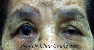
My colleague Dr Ngo showed me this interesting piece of news in a local Chinese newspaper about a Hong Kong woman who shed black tear.
全球第3宗眼皮发霉 港女流黑泪
(香港26日综合电)香港出现全球第3宗罕见流黑色眼泪个案。眼科医院一名44岁女病人,眼睛受到会分泌黑色素的霉菌感染,导致眼皮底下发霉,黑色的霉菌群随泪水流出,变成黑眼泪,并有十多粒黑色的霉菌积聚物,最大一粒近两毫米
I think the picture in the newspaper (shown above) is an artist impression. I can't help noticing that the patients have parallel skin creases (double eyelids) which don't join at the nasal part of the eyes ie not a tapering type of double eyelid (will talk about this later in later blog) typical of Chinese with double eyelids. This type of double eyelids are uncommon amongst Chinese and are most often surgically created or one of the parents is non-Chinese (may be Caucasian, Indian or any South-East Asian natives).
Anyway, I am wondering what the condition was and decided to google under "Hong kong woman black tear" However, there was no news under these keywords but on the web search the newspaper report was actually taken from Archives of Ophthalmology. And the full report is as follow:
Subconjunctival Mycetoma as an Unusual Cause of Tears With Black Deposits
Ocular mycosis is a rare condition that is usually related to ocular trauma, preexisting ocular disease, or immunocompromised states. We report a case of subconjunctival mycetoma secondary to Exophiala dermatitidis in a healthy middle-aged woman with recalcitrant ocular inflammation and black deposits in her tears.
Report of a Case.
A 44-year-old woman had recurrent discharge from her right eye and black deposits in her tears for 2 years. Her symptoms persisted despite the use of topical antibiotics, steroids, and antihistamine. She was otherwise healthy and was not receiving any systemic or other topical medication. She denied any history of ocular trauma or surgery. She did not use contact lenses or eye makeup. On examination, her general condition was excellent. Her visual acuity, intraocular pressure, and fundi were all normal. There was no eyelid swelling or erythema.Oneverting the right upper eyelid, some subconjunctival black deposits were noted . During biopsy, the conjunctiva was incised and multiple black, mulberry-like concretions extruded with mucoid discharge. Topical chloramphenicol, 0.5%, with dexamethasone sodium phosphate, 0.1%, eyedrops were prescribed postoperatively. Histopathological evaluation of these concretions showed large amounts of fungal hyphae with chronic inflammation over the conjunctiva. The diagnosis was subconjunctival mycetoma. Initial culture results for fungal growth were negative, but further evaluation with 28S ribosomalRNAgene sequencing identified the causative organism as E dermatitidis. At subsequent follow-up visits, the patient had complete resolution of symptoms. Topical antifungal treatment was not given as she was asymptomatic and there was no recurrence of mycetoma at month 3 after de´bridement.
Comment.
Tears with black deposits are extremely rare. In our case, we initially thought the black deposits were either foreign bodies or adrenochrome deposits, but they proved to be shedding from the subconjunctival mycetoma. Patients with tears with black deposits should therefore be evaluated for the presence of subconjunctival mycetoma. Asimilar clinical entity termed melanodacryorrhea (black tears) is caused by extraocular extension of uveal melanoma.
In immunocompetent subjects, fungal infection can remain superficial and localized as illustrated in our case. Subconjunctival mycetoma has been reported after subtenon corticosteroid injection in an immunocompromised host and in an immunocompetent woman with no risk factors, similar to our patient. The Exophiala species are dematiaceous mold commonly recovered from soil, plants, water, and decaying wood materials. This strain of black yeasts has been reported to cause deep infection (especially in the lung), cutaneous infection involving skin and mucous membranes, and subcutaneous infection manifested as mycetoma.4 E dermatitidis has been described as the causative agent in fungal keratitis that occurred after keratoplasty5 and laser in situ keratomileusis, 6 but to our knowledge it has not been reported to cause subconjunctival mycetoma.
Treatments described for subconjunctival mycetoma are diverse, ranging from aggressive topical and systemic antifungal treatments following surgical intervention2 to surgical de´bridement alone. A study by Zeng et al4 evaluated the activity of amphotericin B, itraconazole, voriconazole, and posaconazole against E dermatitidis and reported that all 4 antifungal agents have low minimum inhibitory concentrations (range, 0.03-0.5). However, data on correlation between in vitro and in vivo susceptibility are unavailable.
In summary, we describe an immunocompetent woman with tears with black deposits caused by subconjunctival mycetoma, the causative fungus having been identified as E dermatitidis.
Emmy Y. Li, MRCSEd
Hunter K. Yuen, MRCSEd
David C. Lung, MRCPCH (UK)


