Saturday, January 2, 2016

Doc, can you do something about my weird looking eye?

This patient underwent bilateral blepharoplasty and right ptosis surgery a few months ago. She complained the right eye appearing weird and had difficulty opening the left eyelid.


Examination showed that the right eye had poor contour with the highest point of the upper eyelid too far to the temporal side. In addition, there was slight droopy of the eyelid nasally.

The high point should be at the site of red arrow but in this patient, 
the highest point is at the white arrow

In Chinese eye, the highest point of the upper eyelid is usually just temporal to the pupil (in Western eye, the highest point corresponds to the centre of the pupil).  The patient's left eye also had undiagnosed ptosis which was not corrected. 
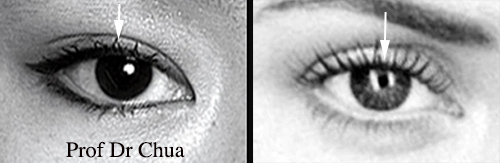
A Chinese eye (right) versus a Caucasian eye showing the highest point 
of the eyelid in Chinese is slightly temporal to the pupil. 

To correct the poor contour, the nasal side of the eyelid had to be lifted. This was achieved by by posterior approach conjunctivomullerectomy. The left ptosis was also corrected the same time using the same posterior approach. 


Before the operation, makings (in blue) were make for anchoring the sutures 
so that the highest point was just temporal to the pupil.

Steps showing the procedure for the right eye. a. The right eyelid was everted; 
b. a horizontal cut was made at the upper border of the tarsal plate, 
the cut corresponded to the distance between the two markings; 
c. Scissors were used to make vertical cuts to produce a strip of 10mm 
conjunctivomuller flap; d. the strip was cut and e. 2 nylon sutures were used to attached 
the cut ends to anterior surface of the tarsal plate; f. at the end of the procedure, 
the highest point has now been shifted nasally.

At the end of the operation, the left ptosis was also corrected.


Friday, January 1, 2016

Doctor, can you remove these lumpy things from my eye?

This young man presented with these lumpy lesions of his right eye for the past 3 months. He has had similar condition 6 months earlier and consulted a different eye doctor. The lesions were excised at that time without further treatment and they returned with a vengeance. 

Conjunctival papillomas affecting the upper and lower tarsal conjunctiva.

Examination revealed multiple pendunculated lesions arising from the upper and lower tarsal conjunctiva and the patient had problem shutting the eye properly. The lesions are typical of conjunctival papillomas. These lesions are benign and are associated with huma papillomavirus (HPV) infection. Although they can be excised easily, recurrence is common unless adjunctive therapy was performed. 

In this patient, the lesions were excised followed by application of 0.01% of mitomycin to the bases for one minute. So far, the patient showed no recurrence at follow-ups. 

The upper eyelid was everted with Desmarres retractor to fully 
expose the affected tarsal conjunctiva.

The lesions were excised using a blade and the bases scrapped clean.

Cotton bud dipped in 0.01% mitomycin was applied to the bases 
for one minute.

At follow-up, there was no recurrence.

At follow-up, the tarsal conjunctiva appeared free of papilloma.


Reference:
Conjunctival papilloma: features and outcomes based on age at initial examination. Kaliki S, Arepalli S, Shields CL, Klein K, Sun H, Hysenj E, Lally SE, Shields JA. JAMA Ophthalmol. 2013 May;131(5):585-93.

Friday, December 18, 2015

Launch of New Book for Sarawak Heart Foundation

I am pleased to announce the publication of the new book "Picture Diagnostic Tests for Postgraduate Medicine" today. The book was written with the help of the Ophthalmology and the Cardiac Department and published by my company Marudi Publication. All proceeds from the sale of the book will be donated to the Sarawak Heart Foundation.

  
Lauching the book with Prof Sim (right) and Dr Koh (left).


The book is now available from the Sarawak Heart Foundation
 in the Heart Centre.

Wednesday, October 14, 2015

Excision and reconstruction of a Lower Lid Lesion

This man was concerned about his lower eyelid xanthelasma which had troubled him for the past 10 years and had got bigger. He had tried trichloroacetic acid and laser treatment but without success. The lesion was excised closed to the margin and a rhomboid flap was used to cover the defect.
(Special thanks to Dr Ho Shu Fen, Dr Chin Ong and Dr Andy Yew for assisting the operations and taking the pictures).
Steps showing the excision of the xanthelasma and use of rhomboid flap to cover the defect. The main concerns in this procedures are lower lid ectropion (eversion of lower lid) and scarring. The former requires the use of flap that does not cause too much tension on the lower lid margin and the later needs meticulous suturing technique to minimal scarring. 

Thursday, October 1, 2015

Excisional Biopsy of a Lower Eyelid Basal Cell Carcinoma

This patient had a lower eyelid ulcer for the past year. The appearance was typical of basal cell carinoma, an excisional biopsy was done with 3mm clear margin. A rotational flap was used to cover the defect as shown. 

Tuesday, August 4, 2015

Doc, what is this in my lower eyelid?

This 70 year-old woman presented with a rapidly enlarging lesion in her right lower lid. The lesion bled easily whenever she rubbed her eye. Examination revealed an ulcerated lesion involving almost half of her right lower eyelid and there was out-turning of the eyelid (ectropion) due to scarring. 

Figure 1. Right lower eyelid basal cell carcinoma with ectropion. 

The appearance was consistent with basal cell carcinoma, a type of skin lesion that typically occurred in sun-exposed skin such as the face and hands. Although cancerous, this type of skin cancer does not spread to distant part of the body. However, if left untreated, it will slowly destroy the surrounding tissues. 
Figure 2. A patient with extensive basal cell carcinoma destroying 
most of the right eyelid, the tumour also invaded deeply. Exenteration 
in which all the ocular tissue were removed leaving behind the 
bony orbit had to be performed. 

The best treatment option is excision with some clear margin to ensure complete excision. Because the lesion in this patient was large, the excision required extensive reconstruction. In this patient, the reconstruction was done using rotation flaps as shown in the photos below.

Figure 3. a. Marking of the lesion with 3mm clear margin; 
b and c. Complete excision of the lesion; 
d. the posterior lamellar was replaced with tarsal plate from the upper lid; 
e and f; the tarsal plate of the upper lid was rotated to the lower lid; 
g. the tarsal plate was sutured to the cut edge of the lower lid; 
h. the anterior lamellar was replaced with upper lid myocutaneous graft; 
i. the upper myocutaneous flap was rotated to the lower lid defect; 
j. to avoid flap retraction the cheek was elevated and sutured to the lateral orbital rim; 
k. the rotation flap was sutured in placed and the harvested area was closed as in upper blepharoplasty; 
l. the right eye was tightly packed for 24 hours to prevent haematoma beneath the flap. 

Wednesday, July 29, 2015

Doc, why do I have so many lines along my double eyelid?

This 36 year-old woman with a history of allergy underwent double eyelid (Asian blepharoplasty) 2 years ago. Initially the double eyelids in both eyes were symmetrical and well-defined. However, in the last 6 months, she noticed the appearance of extra lines in her right upper eyelids which got worse over time (see Figure 1). History revealed she often rubbed her eyelids due to itchiness.

Figure 1. Poorly defined double eyelid 
in the right upper eyelid with extra lines. 

The lost double eyelid (skin crease) was caused by dehiscence (loss of attachment) between the skin and the underlying tissue mainly the levator aponeurosis (the muscle that's responsible for opening the eye). The dehisence was likely to arise from constant eyelid rubbing. Indentation of the eyelid skin showed that the double eyelid can be restored and the extra lines eliminated (Figure 2).

Figure 2. The top picture shows the right eye had well-defined double eyelid
(white arrow) which disappeared towards the corner of the eye (black arrow). 
Indentation of the skin using a paper clip shows the double eyelid 
can be restored with elimination of the extra line. 

There are many ways of correcting this problem. However, the easiest way with a short down-time is to indent the skin with a non-absorbable suture (Figure 3). The principle is similar to the suture technique for Asian blepharoplasty. 

Figure 3. Steps showing how the skin crease was restored. 
a. two marks are made along the original double eyelid near the corner of the eye; 
b. stab incisions were made at the sites marked; 
c. a non-absorbable suture was passed deep through the incisions catching the tarsal plate; 
d. the suture was then passed back through the incision this time just below the skin; 
e. the two ends of the suture were tied; 
f. at the end of the procedure.

At the end of the procedure, there was some puffiness. However, the patient was able to return to work. The recovery could be sped up with ice compress. 

Figure 4. Picture taken one hour after the procedure showing 
well-defined double eyelid despite the swelling.