Thursday, November 18, 2010

Glad that Children don't Have to Suffer Like That

Retinoblastoma is the most common tumour of the eye in children. It has an incidence of 4 per million population. With a population of 27 millions, one can expect 108 cases per year in Malaysia. Most of the cases were picked up during routine eye screening and the most common sign is the presence of white pupil (leucocoria). However, in places where parents don't have easy access to basic health screening, retinoblastoma can develop into disfiguring tumour pushing the eye out of the eye socket. The most common cause of death is spread of the tumour into the brain. Fortunately, with chemotherapy and radiotherapy most patients have a good chance of survival.
Here are some photos and illustrations of retinoblastomas from the 19th century textbooks. Unfortunately, these presentations can still be seen in many developing countries if you google retinoblastoma.

Illustrations of a child with retinoblastoma while alive. The tumour had spreaded
to the skull and brain. The picture on the right was drawn after the child
died and the skin removed.

Pictures of a child with retinoblastom from 1885.

The same child after operation but no front picture was
shown. Without chemotherapy and radiotherapy, the child
was likely to succumb to the disease within a short time even
if the tumour were removed.

In Malaysia, such advanced presentation are rarely seen. This child from a poor family and lived in remote area presented wtih a left swollen blind eye. Although the social worker arranged for the patient to be treated in Sarawak General Hospital, Kuching, the family repeatedly defaulted until the left eye became very swollen. By the time, we saw the patient the left eye was totally destroyed by the tumour and the tumour occupied the whole eye socket. The patient underwent chemotherapy in the children cancer centre and within a month, the tumour shrunk to nothing. Repeated follow up and regular brain scan are needed to detect any early recurrence.
Left eye swollen and painful caused by retinoblastoma.

Tumour and the eye destroyed by the retinoblastoma
shrunk following chemotherapy.

Unfortunately, this tumour is likely to be with us as it is caused by genetic defects. Patients who survive the tumours can pass the genes on to their next generation. So there is a possibility that we will see more and more of this tumour in the year to come as more and more patients survive. This reminds me of an old English consultant whom I used to work for in the UK. He was a Germanophile and appeared to be a strong supporter of the Third Reich. Whenever, he saw a family with two children having this tumour. He would say "If Hitler were around, these people would not be allowed to reproduce!" He was nicknamed Adolf by the clinic nurses ;).

Tuesday, November 16, 2010

How the Excess Upper Eyelid (Dermatochalasis) was Removed

Excess skin of the upper eyelids (dermatochalasis) is a common ageing process. It makes a person appears dull and in severe cases can interfere with vision. The practice of removing the excess skin has been recorded in Egyptian papyrus 3000 years ago. However, with the lack of anaesthesia the procedure was very painful and few patients could bear to go under the knives. One way of overcoming the pain was illustrated in the book Ophthalmodouleia by the famous German eye surgeon Georg Bartisch (1535–1607). In this method, the excess skin was clamped between two rods until the skin died and dropped off which usually took a few weeks.


In the book, the process was described: "The excess skin to be removed is put through the screw clamp and clamped. St. John's wort oil is applied one or twice a day to help the skin comes off quicker and heals. The clamp will fal off in three to four weeks time."

Fortunately we not longer need to use this laborious method. Below show the steps I used to remove excess upper eyelid skin and a few post-operative patients.

This woman has excess upper eyelid skin making her appears tired.
Before the operation, the excess skin is measured with a ruler.

The height of the double eyelid (skin crease) is measured and 
shown to the patient with a mirror. 

The amount of skin to be removed and the height of the
double eyelids (skin creases) are marked.

The excess skin togther with some muscles and fat are removed.

The amout of skin, muscles and fat removed should be the same
on both sides. The double eyelids (skin creases) should be
assessed constantly during operation to make sure they
are of the same heights.

After making sure the heights of the double eyelids (skin creases) are
the same, the skin is stitched up. Firm patching is applied to reduce
post-operative swelling.

Appearance of the eye the second day after operation and
one week later.

Here are three more patients who had had upper eyelid exess skin removed. The procedure not only makes them see better but looks younger.

This patient had upper and lower eyelid excess skin were removed.
Before and 2 weeks after operation.

This patient appears more friendly after the procedure.
Before and 3 weeks after operation.

This patient sees better without having to constantly lift his head.
Before and 3 weeks after operation.

Sunday, November 14, 2010

Just Because it is Not Cancerous

This 60 year-old woman had had this facial blemishes since 5 years ago. She went to her local  polyclinic and was told by the doctor they were not cancerous and she needed nothing done. However, the lesions were causing her a lot of mental distress not least because her grandchildren found them grotesque and refused to be seen with her in the public. She went back to the polyclinic and asked to be referred to a specialist but was told that any removal would result in severe facial scarrings which put her off. Eventually, she decided to put up with them and rarely ventured outdoor. She came to my eye clinic in Sarawak General Hospital two years ago because she developed diabetes and needed eye screening. She was very conscious of her facial lesions so I offered her surgical removal. At first she declined, saying that the previous doctor warned her about the risk of severe facial disfigurements if she were to undergo surgery. Eventually,the desire to appar normal in the public took the better of her and she agreed to undergo surgical removal.

Multiple warty lesios of the face.

Immediate post-opeative appearance of the face.

Appearance of the face two years later. Some lumpy skin
lesions remained but she was happy to leave them.

I excised the lesions below the eyelid and sutured the cut edges. The scar would not be obvious as it follows the natural line of the skin (relaxed skin-tension lines as shown in the picture below). The lesions around the mouth were shaved superficially instead of excisions so that they would not form obvious suture marks. The patient was extremely pleased with the result and the first thing she did was to change her hair style and bought some new clothes. I am pleased that her confidence was restored and she not longer became the centre of attention whenever she appeared in public or shun by her grandchildren.
Diagrams showing the relaxed skin-tension lines.
Incision along these lines creates minimal scarring.


Lesson of the story:
1. Facial blemishes can severely affect the patient's confidence even if it is not cancerous.
2. Doctors can discouraged the patients from operation if they exaggerate the post-operative complications.

Friday, November 12, 2010

How they Used to Torture the Nun

Abscess of the tear (lacrimal) sac is a painful condition and caused by blockage of the nasolacrimal duct (see picture below) which connects the sac to the nose. This unfortunate Austrian nun of the 17th century had this affiliation and was treated surgically without anaesthesia (which had yet to be discovered). She was tied up in the chair and restrained by other nuns whie a priest with a sharp knife cut the abscess to drain it.

A print from a 17th century Austrian medical textbook.

A close-up of the nun after the incision


Fortunately, the same condition can now be treated humanely. Here are two cases which I did recently. The first one had an external dacryocystorhinostomy (DCR) in which a connection is made between the lacrimal sac and the nasal cavity so that the blockage is by-passed. 

This patient had chronic mucous discharge due to blocked duct.
A mark is made where skin would be made to create a connection
between the lacrimal sac and the nasal cavity.

Syringing of the lacrimal sac caused the mucous discharge
to enter the eye.

The skin is incised and the muscles pushed aside to reveal
the bone (white in colour).

The nasal bone is removed with a bone crusher.

A hole is made in the nasal bone to reveal the nasal mucosa.

A cut is made in the nasal mucosa to form a flap.

The lacrimal (tear) sac is cut to form flap.

A tube is inserted to keep the passage patent.

The tube is pulled though the nose and tied.

The flaps from the nasal cavity and the lacrimal sac
are stitched together,

The skin incision is sutured.

The illustration summarizes the procedure above.

The second one had the sac removed. The patient had a seious road traffic accident 5 years ago resulting in lost of the right eye and multiple facial bone fractures. There was recurrent abscess formation which was incised and drained. As the patient had complex orbital and nasal bone fractures, I decided not to perform dacryocystorhinostomy (DCR) and instead remove the entire sac.

Before and one week after excision of the tear sac.


Thursday, November 11, 2010

What the Irish can Offer You and it is not Their Bonds

This day the celtic tiger is turning into PIIGS and nobody wants to touch the Irish bonds with a barge pole. However, there is one thing not self-respecting middle-aged women and men can do without which is found only in the West Coast of Ireland – BOTOX!. The Allergan factory that produces the entire wold supply of Botox is located in the picturesque town of Westport in County Mayo.
The local claims the Westport scenery is as good in erasing
the frown lines as their famous product.

Botox is the most lethal toxin known to men and the cause of food poisoning (botulinism). The bacteria that produce it was discovered in 1895. However, it was not until 1950s that scientists began to experiment with botox medically and found that in minute quantity it can paralysed selective muscles. Until the late 1980s, botox was only used for crossed eyes (strabismus) and uncontrolled blinking or facial twitching (blepharoplasm and hemifacial spasm).

The use of it as wrinkle erasers was discovered by chance by a couple of husband and wife ophthalmologist in Vancouver. Having a sharp eye, the wife noticed that patients treated by her husband for uncontrolled blinking had less wrinkles. As they say the rest is history, the couples are now Hollywood doctors to the rich and famous. Just goes to show if you open your eyes, they are fortunes to be made. This is good news for my junior doctors who helps me run a 3-monthly Botox clinic for over 40 patients with blepharospasm and hemifacial spasm from all over Sarawak.
Alaistair and Jean Carruthers, the husband and wife ophthalmologists
who are the saviours of middle-aged women all over the world.

Aside from erasing the wrinkles, lifting the eyebrows, giving ones a good jaw line, there are many less glamorous uses of Botox including hyperhydrosis (excessive sweating of the arm-pit), spasticity of arms and legs in cerebral palsy and after strokes. More recently it has been approved for migraine.
Lesson of the story: “Dump Irish Bonds, buy Allergan Shares”


Wednesday, November 10, 2010

I am too Sexy for My Passport

After eyelid surgery, some patients prefer to take a long holiday away from friends so that upon their return there was few signs left to suggest the lids had been operated. Usually, this is no a problem if one travels within Malaysia but for those who wish to travel to countries where visa are acquired the newly acquired eyelids can cause problem as their facial features can differ greatly from their passport photos.
This old man underwent upper eyelid surgery to remove the excess skin that covered his vision. After the operation, he decided to travel to China for a long break, however, the embassy refused to issue him a visa as his face did not match that of his passport. The visa was eventually issued after he obtained a medical report from my clinic stating he had had eyelid surgery. However, on the way out of China he was once again detained at the custom for suspected identitiy theft.

Before and after eyelid surgery.

I now routinely advise patients who have significant post-operative alterations in their facial appearance to change their passport photos as soon as possible to avoid problems at the customs. Otherwise, a holiday in Genting Highland is as good and I don't say this just because I have shares in Genting Casino.

Monday, November 8, 2010

Shxx! My Discounted "Sheep Placenta" Injection Turned Out to be .............

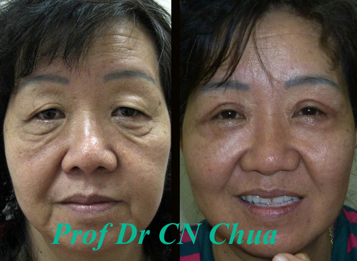
This woman was a go-between for an unlicensed travelling doctor from China who specialized in "Sheep Placenta" injection amongst other things. She was impressed that patents who had had cheek injection of the "Sheep Placenta" immediately achieve wonderful contours. The injections were performed either in some hotel rooms or the doctor would pay home visits. After seeing the miraculous results, she decided to have a go and asked for free injection but the doctor only agreed 50% discount saying "My dear, this "Sheep Placenta" is extremely expansive and comes all the way from Switzerland. I only agree to give you 50% discount because you are such as a good go-between in bringing me patients. If all my go-betweens get free treatments, I would go bankrupt!" Thinking that she got a good deal, she could not wait to have the injections to get her new cheeks.


While her cheeks looked good in the first year, she started to notice something not quite right as the injected areas became harder with time and appeared to grow in size. She came to see me for eyebag removal. However, the swollen areas appeared hard and not mobile suggesting some foreign particles had been injected. She desperatedly wanted the things taken out so I performed lower eyelid surgery (blepharoplasty) and removed the hard particles. These were sent for analysis and the pathologist reported these as foreign materials most likely to be from silicone injection.
Hardened and swollen cheeks from the injection.

Hardened areas excised as hard masses firmly attached
to the skin and muscles.

Histology showed chronic inflammation consistent
with silicone injection.

Silicone injection is illegal because it can cause tissue distortion as a result of chronic inflammation. As the distortion may take more than a year to develop, the "doctors" can conveniently deny the distortions are the result of their injections. Unfortunately, silicone injection is used widely in many nonlicensed aesthetic centres and packaged as "Sheep Placenta". It is not surpising as the profit margin is very high. A bucket of silicone costs a few hundred ringgits and the operators can make hundred of injections out of it charging anything from a few hundreds to a few thousands each session.

In a way, this woman had it easy as she was only given injectioins to the cheek. I dread to think of those gullible women that had had full facial treatment. Some of them may well end up like these unfortunate women.

Distorted and lumpy areas as a results of previous
silicone injections.

The following is warning from FDA (USA Food and Drug Adminstration) "the marketing of liquid silicone for injection for any cosmetic purpose, including the treatment of facial defects or wrinkles, or enlarging the breasts are not approved. The adverse effects of liquid silicone injections have included movement of the silicone to other parts of the body, inflammation and discoloration of surrounding tissues, and the formation of granulomas (nodules of granulated, inflamed tissue)."

Moral of the Lesson: "Don't receive any injection from unqualified personnels even if it is discounted and do not introduce your friends to them either!"